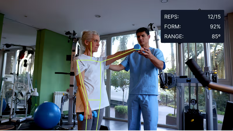
Patient Positioning AI

Position every patient markerlessly, cut retakes on CT, MRI, X-ray, and mammography, and add markerless SGRT and DIBH gating on every linac, on the depth and RGB cameras your suites already run.
Shipping container with OCR output

Patient Positioning AI Across Radiology, Radiation Therapy, and Every Modality

Deploy Anywhere, Run Everywhere

Run patient positioning on CT and MRI gantry cameras, linac SGRT depth cameras, X-ray and mammography suite cameras, PET/SPECT, and interventional radiology, on the edge, on-prem, in a hospital-controlled VPC, or air-gapped, wherever your suite needs it. Camera-agnostic, no proprietary positioning hardware required.

One Platform, Full Adoption

Tools every hospital team can adopt, from radiologic technologists, mammographers, and radiation therapists to medical physicists, radiation oncologists, biomedical engineering, and hospital leaders owning throughput and HCAHPS, no separate ML team required to ship and own positioning models.

HIPAA-Ready, Audit-Ready, Reimbursement-Ready

Data stays safe with HIPAA compliance, SOC 2 Type II, encrypted data, hospital-controlled deployment, and an uptime SLA on every modality. Audit-ready records support FDA 510(k), IEC 60601-1, IEC 62366, ISO 13485, DICOM (including DICOM RT), HL7 v2 and FHIR, IHE Radiology profiles, ACR accreditation, NRC/IAEA radiation therapy standards, ASTRO clinical practice guidelines, CMS documentation, and Joint Commission survey requirements.
Markerless SGRT & DIBH Gating on Every Linac
CT, MRI & X-Ray Retake Reduction
ISO Center & Anatomic Landmark Detection
Patient ID & Wristband OCR (HIPAA)
Mammography, PET/SPECT & Interventional
DICOM, HL7, FHIR, PACS, EMR & TPS Integration
Markerless SGRT & DIBH Gating on Every Linac
CT, MRI & X-Ray Retake Reduction
ISO Center & Anatomic Landmark Detection
Patient ID & Wristband OCR (HIPAA)
Mammography, PET/SPECT & Interventional
DICOM, HL7, FHIR, PACS, EMR & TPS Integration
Markerless SGRT & DIBH Gating on Every Linac
CT, MRI & X-Ray Retake Reduction
ISO Center & Anatomic Landmark Detection
Patient ID & Wristband OCR (HIPAA)
Mammography, PET/SPECT & Interventional
DICOM, HL7, FHIR, PACS, EMR & TPS Integration
Markerless SGRT & DIBH Gating on Every Linac
CT, MRI & X-Ray Retake Reduction
ISO Center & Anatomic Landmark Detection
Patient ID & Wristband OCR (HIPAA)
Mammography, PET/SPECT & Interventional
DICOM, HL7, FHIR, PACS, EMR & TPS Integration

Talk to a Vision AI engineer who's shipped patient positioning in radiology and radiation therapy.

A single mispositioned patient on a CT chest scan, a missed ISO center at treatment planning, or a repeat mammography compression can mean a retake that doubles patient radiation dose, a linear accelerator fraction that misses the tumor and hits organs at risk, or an HCAHPS hit that costs the hospital reimbursement points. Bring us your toughest patient positioning problem and we'll map a working solution.
  • Solution architecture for HIPAA, FDA 510(k), IEC 60601-1, ISO 13485, DICOM, HL7 v2/FHIR, IHE Radiology profiles, ACR accreditation, NRC/IAEA, and Joint Commission survey documentation
  • Live demo on your CT gantry, MRI suite, linac SGRT camera, mammography, X-ray, PET/SPECT, or interventional footage with de-identified imagery
  • Deployment options: edge, on-prem, hospital-controlled VPC, or air-gapped, with integration into PACS, EMR/RIS, TPS, and oncology information systems through DICOM, HL7 v2, FHIR, and IHE profiles
  • ROI modeling against retake radiation dose, scanner-hour throughput loss, linac fraction accuracy, HCAHPS impact, staff musculoskeletal load, and payer medical-necessity documentation
We will connect you with an AI subject matter expert on our team based on your answers.
What challenges would you like to solve with vision AI?
Where will you run vision AI?
Are you replacing a current solution with AI or will this be a new solution?
How many detections do you anticipate per month?
Describe the business problem you would like to solve.
Oops! Something went wrong while submitting the form.

Over 16,000 organizations build with Roboflow.

“Roboflow has been instrumental in accelerating our learning and deployment of innovative AI solutions”
Travis Turnbull Vice President & CIO, Pella Corporation
See customer stories
Trusted by top manufacturers

Bring Real-Time Intelligence to Every Patient on Every Scanner and Every Linac, with Vision AI

Add a real-time positioning layer to every patient on every modality with Vision AI for patient positioning. Built for the hospitals where one CT chest retake means eight extra chest X-rays worth of dose to a patient who already came in sick, one linac fraction that misses the tumor and hits an organ at risk hurts a real patient, one repeat mammography compression is one more painful compression, one missed patient ID at scanner setup can mean the wrong study on the wrong record, and one throughput hit at the busiest CT cascades into HCAHPS complaints and schedule slippage. Whether you're positioning patients for CT, MRI, X-ray, digital mammography, fluoroscopy, PET/SPECT, interventional radiology, or radiation therapy on linear accelerators, Roboflow extends positioning coverage to every scan and every treatment fraction, on the depth and RGB cameras your suites already run.

Radiology Positioning: CT, MRI, X-Ray, Mammography, PET/SPECT:

  • Detect patient landmarks (head, sternum, ISO center, anatomic markers) markerlessly with RGB, depth, and structured-light cameras, and align to the modality's planned protocol before the first exposure
  • Cut retakes on CT (5-15% historical), X-ray (15-25%), and mammography (5-10% recompression), reducing patient dose, staff musculoskeletal load, and scanner-hour throughput loss that hits HCAHPS
  • Coach patient motion in real time (breath-hold prompts, positional-drift alerts, comfort adjustments) with the technologist in the loop and the patient in view of the console

Radiation Therapy: Markerless SGRT, DIBH Gating, and ISO Alignment:

  • Add markerless surface-guided radiation therapy (SGRT) on any linac (Varian TrueBeam, Elekta Versa HD, Elekta Unity MR-linac, ViewRay MRIdian), with sub-millimeter surface tracking against the planned reference from the planning CT and TPS
  • Coach and gate deep-inspiration breath-hold (DIBH) for left breast SBRT and other cardiac-sparing treatments, with a beam-gating signal the linac controller can act on if the patient drifts out of tolerance
  • Reconcile positioning against the TPS (Varian Eclipse, Elekta Monaco, RaySearch RayStation, Brainlab Elements) and push per-fraction logs into the oncology information system (Varian ARIA, Elekta Mosaiq) via HL7 and IHE RO profiles

Patient ID, Workflow Integration, and Compliance:

  • Verify patient identity at modality setup with wristband OCR and opt-in facial-biometric confirmation (HIPAA-compliant, hospital-controlled), tying the physical patient to the order in the EMR/RIS (Epic Radiant, Cerner Millennium, Meditech Expanse) and preventing wrong-patient wrong-study events
  • Integrate with PACS (Sectra, Merge/Change Healthcare, Agfa, Fujifilm Synapse, Philips IntelliSpace, GE Centricity) via DICOM, EMR/RIS via HL7 v2 and FHIR, and radiation oncology systems via IHE RO profiles (RO-ILS, RO-SWF), with REST, MQTT, and OPC UA for hospital IT
  • Deploy on-prem, on the edge at each scanner or linac, in a hospital-controlled VPC, or air-gapped, with SOC 2 Type II, HIPAA, encrypted data, and audit-ready records for FDA 510(k), IEC 60601-1, IEC 62366, ISO 13485, ACR accreditation, NRC/IAEA, ASTRO, CMS documentation, and Joint Commission surveys

Bring intelligence to every patient today. Stop retakes, mispositions, and wrong-patient events from becoming radiation dose to the patient, linac fractions that miss the tumor, HCAHPS complaints, or HIPAA reportable events.

More About Patient Positioning

What is patient positioning with Vision AI?

Patient positioning with Vision AI uses computer vision models on RGB, depth, and time-of-flight cameras to detect patient landmarks (head, sternum, ISO center), track patient surface in real time against a planned reference from the planning CT and TPS, coach breath-hold and motion management, verify patient identity with wristband OCR, and reconcile positioning across CT, MRI, X-ray, mammography, PET/SPECT, interventional radiology, and radiation therapy on linear accelerators. The system integrates with PACS, EMR/RIS (Epic, Cerner, Meditech), TPS (Varian Eclipse, Elekta Monaco, RaySearch RayStation), and oncology information systems (Varian ARIA, Elekta Mosaiq) via DICOM, HL7 v2, FHIR, and IHE Radiology profiles. Hospitals, imaging centers, and radiation oncology practices use it to cut retakes and radiation dose, add markerless SGRT and DIBH gating, prevent wrong-patient wrong-study events, protect HCAHPS scores, and document compliance under HIPAA, FDA 510(k), IEC 60601-1, ISO 13485, ACR accreditation, NRC/IAEA, ASTRO, CMS, and Joint Commission surveys.

Can Vision AI hit sub-millimeter accuracy for markerless SGRT and DIBH gating?

Yes. Sub-millimeter markerless SGRT and DIBH gating on left breast SBRT is exactly where radiation oncology feels the most pressure, because DIBH spares the heart and LAD by pulling breast tissue off the chest wall, and a patient who drifts out of the breath-hold window during beam-on delivers dose to cardiac tissue rather than the tumor. Roboflow continuously reconstructs a patient-surface point cloud from depth and structured-light cameras above the couch, aligns it to the planned reference surface from the planning CT and TPS at every frame, and exposes a beam-gating signal the linac controller (Varian TrueBeam, Elekta Versa HD) can act on if the patient drifts out of the physicist-defined tolerance (typical 2-5 mm translation, 1-3 degree rotation). The honest tradeoff: a dedicated purpose-built SGRT system remains the reference for the initial commissioning baseline at most clinics, and Roboflow is positioned as the deep-learning positioning layer that scales across CT retake reduction, mammography, MRI patient comfort, wristband OCR, and interventional radiology on the same platform, so the hospital does not maintain a separate vendor stack per modality.

Does patient positioning support HIPAA, FDA 510(k), IEC 60601-1, DICOM, HL7, and ACR accreditation?

Yes. Roboflow models can be trained against your specific HIPAA and HITECH protections, SOC 2 Type II, FDA 510(k) premarket notification, IEC 60601-1 and 60601-1-2 (medical electrical equipment safety and EMC), IEC 62366 (usability engineering), ISO 13485 (medical device QMS), ISO 14971 (risk management), DICOM (including DICOM RT and Structured Reporting), HL7 v2 and FHIR, IHE Radiology profiles (SWF, PIR, XDS-I, RO-ILS, RO-SWF), ACR accreditation for CT, MRI, mammography, and radiation oncology, NRC and IAEA for radiation therapy, ASTRO clinical practice guidelines, CMS reimbursement documentation, and Joint Commission survey requirements. The system logs every positioning event with hashed patient ID, modality, protocol, reference surface, delta from reference, beam-gating events, operator ID, and timestamp against the acceptance criteria your medical physics and quality teams define, and produces audit-ready records for FDA 510(k) design history file evidence, ACR surveys, ASTRO APEx review, and CMS medical necessity documentation.

Can it integrate with our PACS, EMR, RIS, TPS, oncology information system, and hospital IT?

Yes. Roboflow Inference exposes a standard API and supports common hospital and radiology protocols. Customers integrate with PACS (Sectra, Merge/Change Healthcare, Agfa, Fujifilm Synapse, Philips IntelliSpace, GE Centricity, Carestream Vue), EMR/RIS (Epic Radiant, Cerner Millennium, Meditech Expanse, Allscripts), TPS (Varian Eclipse, Elekta Monaco, RaySearch RayStation, Brainlab Elements, Accuray Precision), oncology information systems (Varian ARIA, Elekta Mosaiq), image-guided surgery navigation (Medtronic StealthStation, Brainlab Curve, Stryker Q Guidance), and interventional radiology systems (Philips Azurion, Siemens ARTIS, GE Innova) through DICOM (including DICOM RT), HL7 v2, FHIR, IHE Radiology profiles, REST, MQTT, and OPC UA. Deployments run on-prem in the hospital data center, on the edge next to each scanner or linac, in a hospital-controlled VPC, or air-gapped, with per-event audit-trail records that support FDA 21 CFR Part 11 style attribution, FDA 510(k) design history file evidence, ACR accreditation, ASTRO APEx, and Joint Commission survey documentation.

Stay Connected

Get the Latest in Computer Vision First

Thank you for subscribing!
Oops! Something went wrong!.
Unsubscribe at any time. Review our Privacy Policy.

Additional resources

Vision AI is transforming every industry. Let’s transform yours.